Cervicalgia (Neck pain): causes, symptoms, and remedies in the case of inflamed cervical spine
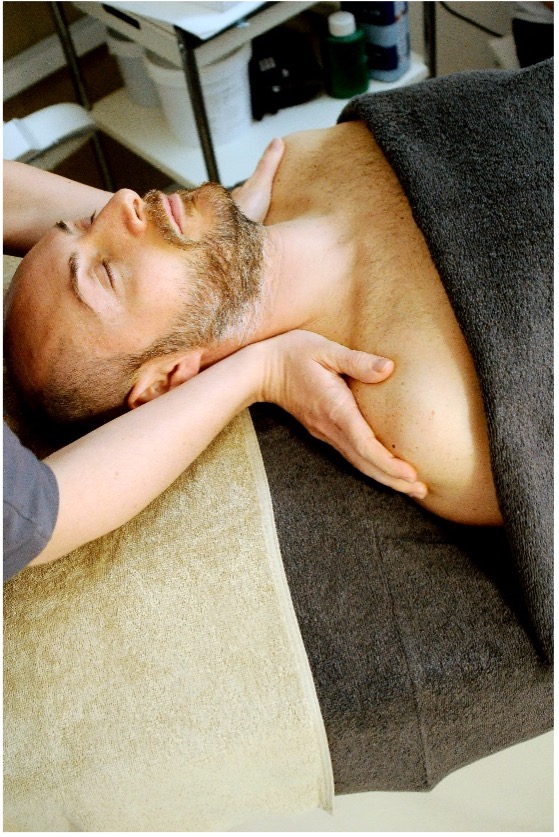
What can I do if I have neck pain or cervical spine problems, and how can I alleviate the pain due to cervical spine issues? Read here for the best exercises, remedies and advice to sleep well.
Neck pain, caused, for example, by an inflamed cervical spine, is one of the most common musculo-skeletal problems for which people seek the help from a doctor or physiotherapist. Although its progress is benign, and acute attacks tend to subside spontaneously, neck pain has serious repercussions in terms of financial and social costs, causing a notable number of days off work. It is also worth noting that 10% of these cases, due to recurrent episodes, tend to become chronic.
The wealth of visceral and nervous elements in the neck, and the ease with which referred pain occurs, require a careful gathering of information aimed at identifying the possible presence of red flags, i.e. disorders not related to muscles or the skeleton, the symptoms of which may include cervical spine (neck) pain.
Cervicalgia (Neck pain): an anatomical view
According to the different anatomy and biomechanics it is a good idea to divide the cervical spine, i.e. the set of vertebrae that make up what is commonly defined as the C-spine, the inflammation of which leads to neck pain, into the following sectors:
– Upper C-spine: occiput-C3
– Middle C-spine: C3-C5
– Lower C-spine: C5-C7
In the Upper C-spine, the architecture is distinguishable above all due to the lack of intervertebral discs and the special shape of the Atlas (C1) and the Axis (epistropheus) (C2). Another distinguishing feature is the orientation of the upper joints of the head in relation to the other facet joints and the complex extent of ligaments.
In the middle/lower C-spine, in addition to the intervertebral discs and facet joints, there are also the uncovertebral joints.
Inflamed cervical spine: what are the symptoms?
In the case of pain due to the C-spine, the symptoms vary in number and type, depending on the level and structures involved. Here is a list of symptoms, but bear in mind that these can vary greatly from patient to patient:
– Upper C-spine: pain in the nape or lower nape area, cervicogenic headache (secondary headache), vertigo and dizziness, tinnitus, pain in the neck through to the upper fascia of the trapezius
– Lower and Middle C-spine: pain the neck, pain in the trapezius, pain between the shoulder blades, pain in the shoulder, fascia pain along the arm, tingling/numbness of upper limbs, muscle weakness, burning or electric shocks.
Possible causes of neck pain
The specific cause is always difficult to identify, as it is often due to several factors, above all in the case of chronic neck pain. We have used the following clinical models as examples:
– Disc pain and cervical herniated disc
– Radicular syndrome
– Dysfunction of the facet joints
– Weakness in deep muscles of the neck or upper back muscles
– Muscle contractures and trigger points
– Referred pain from visceral tension
– Postural problems due to incorrect posture, such as spine distorsion, forward head, posterior muscle retraction, etc.
– Mental and physical stress
– Cervical arthritis
Cervical herniated disc
The cervical disc disorder, or herniated disc, is a frequent reason for visiting the orthopaedic specialist, neurologist, or physiotherapist as neck pain, with or without radiation to the brachial plexus (shoulder and arm), may be the main symptom of a series of serious illnesses. A rehabilitative approach must always be investigated when the clinical and instrumental examinations exclude a paralysing type hernia requiring surgery.
Literature in this regard highlights the importance of a rehabilitative approach based on therapeutic exercise, associated with a diverse range of physical therapies to achieve optimal pain relief (e.g. TECAR or LASER). Indeed there is strong evidence in favour of multi-disciplinary treatments with manipulation or mobilisation, and targeted exercises.
The intervertebral disc is made up of a soft inner core (nucleus pulposus), which cannot be compressed but can be deformed, and on the outside a ring of fibrous cartilage (anulus), which is more solid, anchored to the bone tissue of the vertebral body. Active vascularisation occurs up to the age of 20; then a diffusive mechanism comes into action, which provides for nutrition, and so involution processes can slowly start, towards degenerative disc disease.
The spinal column is very mobile, placed between the rigid rib cage and the heavy head, and is therefore subject to damage by wear and various traumas such as whiplash. As the joints C4-C5, C5-C6 and C6-C7 are those with most mobility, degenerative disc diseases primarily regard these levels.
In the case of herniated disc, the clinical diagnosis comprises a series of symptoms and neurological signs that vary depending on location, type and extent of the hernia.
The symptoms are normally root-related (due to compression and/or irritation of a nerve root), even if, in the case of large medial hernias, it may be associated with bone marrow compression. The most common issues that patients complain of, or which are seen in clinical examinations are:
– Pain: mainly felt in the area of nerve distribution concerned
– Paresthesia: tingling, numbness etc.
– Hypoesthesia: reduction or loss of sensation in the dermatome corresponding to the nerve root concerned
– Vegetative symptoms
– Hyposthenia: varying degrees of loss of strength
– Muscle hypotrophy more or less severe
– Reduction or loss of tendon-bone reflex
Other symptoms include stiffness in the morning or after holding a position for a long time and defensive type muscle spasms.
The diagnostic examination most commonly used to confirm or exclude a herniated disc is the Nuclear Magnetic Resonance (RM) which enables an accurate identification of the location/s and entity of the hernia.
Therapy includes symptomatic treatment and treatment of the cause:
– Rest: advised above all for acute cases
– Collar: without exaggerating, we may advise the use of a collar to offload the cervical segment
– Physiotherapy: the aim of this is to improve the tone and trophism of the muscles in the spinal column and to re-educate vertebrae functionality. This is the most important phase: therapeutic exercise represents the core of the rehabilitation plan.
– Other physical therapies: for example TECAR therapy, combined with massage and mobilisation, can help release muscle spasms and can significantly reduce symptoms.
– Pharmacological treatments: prescribed by the doctor, and may include NSAIDS (non-steroid anti-inflammatory drugs) and/or cortisone.
Remedies for neck pain
In the absence of red flags, the physiotherapist will evaluate and decide on the best treatment to undertake.
The aims in the acute phase (e.g. stiff neck), are rapid reduction of pain and recovery of mobility. In the chronic phase, the main aim is to identify the main cause of the symptoms, to the point of bringing the patient to stable relief of symptoms.
- Manual therapy
- Therapeutic Exercise
- Instrumental therapy
- Postural re-education
- Stress management
- Home exercises
- Educating the patient on what to avoid
Useful exercises for neck pain
This section provides a number of exercises that can be done alone at home in the case of neck pain. If any of the exercises increases rather than reduces pain, stop them immediately and speak to your physiotherapist.
– Self-mobilisation combined with breathing: sitting on a chair with the back straight, breathe out and at the same time turn the neck slowly to the right. Breathing in, slowly return to a central position. Breathing out, turn the neck slowly in the opposite direction. Repeat this for 4’-5’, always remaining below the pain threshold.
The same exercise can be done by bending your head to the right and left (ear to shoulder) or flexing/extending the C-spine (looking up and down).
– Lateral muscle stretching: sitting on a chair with the back straight, grip the right side of the head with your left hand and lightly stretch the neck towards the left shoulder, keeping the right shoulder low and relaxed. Stay below the pain threshold and hold this position for 1’, breathing deeply. Then perform the same exercise on the other side.
– Contraction-release of the Trapezius: sit on a chair with the back straight, and arms relaxed along your sides. From this position, lift your shoulders towards your ears, clenching the trapezium muscles on both sides. Clench for 5’’, after which quickly and completely release both muscles, letting your arms hang on both sides. Repeat 10 times.
More details. Neck pain and sleep: how to sleep well
Let’s start with the position in bed you should never keep if you have a stiff neck: never lie on your stomach with you neck turned to one side. In fact, to get into this position, you need to rotate joints by around 90°. In the case of a stiff neck, getting into this position places demands on our vertebrae that are beyond their ability, and so we then have to compensate in the lumbar region, for example, which could lead to back ache. Also, keeping this position overnight could lead to discogenic wry neck (stiff neck) as described above.
The best lying positions for the spinal column are on your side or on your back, where the choice of pillow is also important for both:
– Lying on your side: the pillow must enable correct alignment of the C-spine with the rest of the spinal column, so it is advisable to choose a pillow that is not too soft, to prevent the neck from bending too much towards the shoulder below, and not too raised to prevent excessive lateral bending towards the ceiling.
– Lying on your back: also in this position you need to check C-spine alignment, avoiding above all that the nape falls too low into the mattress, creating excessive lordosis of the C-spine section. In the same way, a pillow that is too high would mean that the neck bends too much, which is not advisable for intervertebral discs.
Conclusions
C-spine disorders are undoubtedly of significant interest, with a variety of problems as we have seen above, and which are increasingly common.
It is essential to have an initial evaluation by a specialist doctor and physiotherapist so that the best rehabilitation treatment can be decided on, depending on the clinical case in hand. Indeed, rather that thinking of treatment protocols, we need to focus on personalising therapies, where a vital element is the technical know-how of the physiotherapist, who can really make a difference in resolving symptoms and treating the cause of the dysfunction of the C-spine.
Sources:
“Trattato di medicina fisica e riabilitazione” [An essay on physical medicine and rehabilitation”] edited by Valobra, Gatto, Monticone, “Modelli clinici in terapia manuale” [Clinical models in manual therapy”] edited by Westerhuis e Wiesner.
